| First Section Page | Page 14 of 14 | Last Section Page |
Patient Assessment and Site Selection Using Ultrasound
(continued)
Subclavian Vein
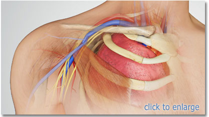
The subclavian vein can be difficult to visualize due to its position beneath the clavicle. However, it is possible to visualize the vessel by placing an endocavitary probe in the supraclavicular fossa, lateral to the clavicular head of the SCM. A long axis view shows the subclavian vein merging with the internal jugular vein, both ultimately forming the innominate vein. Placing the patient's arm behind his or her back improves the visualization.
Femoral Vein
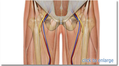
When assessing for femoral placement, distinguish the femoral vein from the femoral artery and femoral nerve. Remember, femoral placement is not recommended for adults unless for emergent use or last possible resort for access. If a femoral catheter is placed, a new catheter at another location should be placed within 24 hours and the femoral catheter removed, when possible.
Go to Section 6:
Step by Step Insertion Techniques ![]()
| |
Page 14 of 14 |