| First Section Page | Page 3 of 14 | Last Section Page |
Ultrasound Theory and Point of Care Application
(continued)
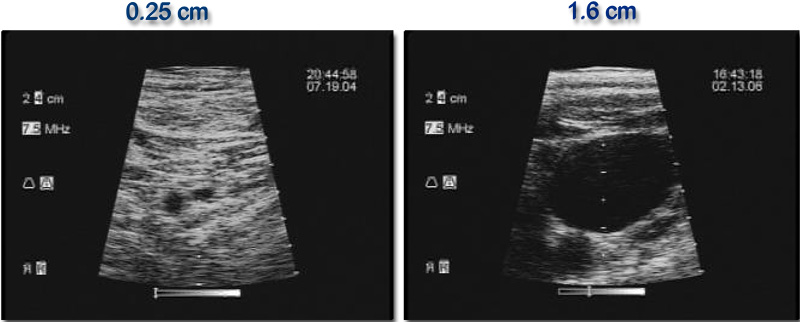
The anatomy of veins and the landmarks that define them are variable and are often impossible to visualize with the naked eye. One prospective study using ultrasound to guide placement found that nearly two-thirds of people have an asymmetry in their internal jugular veins, and the right vein was dominant in less than 70% of cases. Nearly one-quarter of people's veins were 0.4 cm2 or smaller, which can create serious complications (). Other research has found anatomical variations in 8% of internal jugular veins examined with ultrasound (,).
Indeed, millions of CVCs are inserted every year in the United States, and up to 20 percent of cases may report a failure to cannulate the vessel (). When relying on surface landmarks to guide placement, complications occur in 10 percent of cases, and can be significantly higher in pediatric cases (). Additional research suggests the use of ultrasound in CVC placement may reduce the rate of injuries by 25 percent ().
Several randomized controlled studies have compared the use of real-time ultrasound to blind insertion and results consistently show that imaging significantly reduces the failure rate, complication rate and the total number of attempts (). One prospective randomized study of 900 patients undergoing ultrasound guidance for placement of internal jugular vein catheters showed that ultrasound increased the overall success rate to 100 percent (versus 94 percent in blind placement), reduced carotid puncture to one percent (versus 11 percent in blind sticks), reduced the rate of hemothorax and pneumothorax and decreased infections ().
| |
Page 3 of 14 |